*Saiga horns are also known as ling yang (羚羊) and used in Traditional Chinese Medicine
By Richard A. Kock and E.J. Milner-Gulland
Reproduced by kind permission of ‘Natural History‘ magazine. First published in the April 2018 issue.
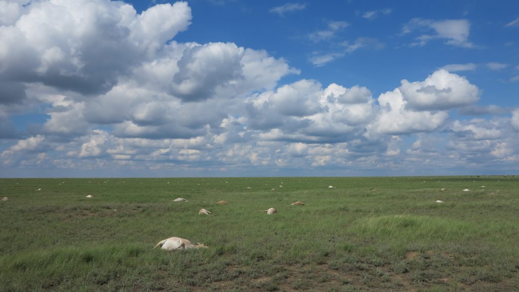
Saiga antelope litter the ground during a mass die-off in May 2015 in the Betpak-dala region of Kazakhstan.
Every year at calving time for the past six years, an international team of researchers—under the authority of the government of Kazakhstan and in collaboration with Kazakhstan research institutions—has been monitoring the health of the saiga antelope population in central Kazakhstan.
Saigas once roamed widely over steppes and semi-arid deserts from southeastern Europe to Mongolia and into China. Now, one population of Saiga tatarica tatarica, the nominate subspecies, exists in Russia, three in Kazakhstan, one of which migrates to Uzbekistan in the winter. There is another distinct sub-species, Saiga tatarica mongolica, in western Mongolia. Populations in the rest of their once-wider range are extinct.
Since 2002, the World Conservation Union’s (IUCN) Red List has listed saigas, Saiga tatarica L., as Critically Endangered. The species was listed as lower risk-conservation dependent only in 2000, but then suffered a rapid, more than ninety percent, decline in its global population, due to poaching after the break-up of the Soviet Union, leading to a population of less than 50,000 in 2003.
As one of us (Milner-Gulland) reported in a 2001 paper in Oryx, the literature shows that “Saiga populations have fluctuated dramatically over the last century, principally as a result of hunting for meat and horns, and climatic variability. The horns are borne only by males and are used in traditional Chinese medicine. Data on historical changes in the numbers and range of the Mongolian subspecies are minimal. The nominate subspecies was heavily hunted in the nineteenth century, and by the time of the Soviet revolution was reduced to a few thousand individuals. A complete ban on hunting allowed populations to recover, and regulated commercial hunting was started in the 1950s. Regulated hunting, principally for meat, continued throughout the Soviet period. Since then, a collapse in funding and infrastructure for saiga management, combined with a disintegrating rural economy, has led to uncontrolled large-scale poaching for meat and horns.”
From 2005 to 2015, some populations of the species started to recover. By 2015, there were more than 300,000 individuals in existence—of which 240,000, or more, were in the Betpak-dala population of Kazakhstan. In Soviet times, Kazakhstan’s saiga populations were well monitored by teams of scientists from the Institute of Zoology of the Kazakhstan Academy of Sciences. After the break-up of the Soviet Union, monitoring became more patchy, due to lack of resources and capacity. However, in the last few years, detailed monitoring of the Betpak-dala population, and to a lesser extent the other populations, has restarted, led by the Kazakhstan government’s Committee on Hunting and Forestry, with scientific input from the local non-government agency, the Association for the Conservation of Biodiversity in Kazakhstan. International veterinary researchers joined this effort, in response to a disease outbreak in the Ural population in 2010. In the Soviet literature, mass deaths from disease have been reported in 1974, 1981, 1988, and 2010. In 1974, the cause was foot-and-mouth disease from livestock (pre-vaccination), while in the other years, various types of pasteurellosis, a bacterial infection, were implicated.
In 2014, calving went without incident. The population appeared healthy and growing. Before the start of the 2015 calving season, aerial surveys determined the locations and sizes of gathering herds. By the start of calving, which happens over a wide area, monitoring teams were in place on the ground, scattered among the three saiga population sites in Kazakhstan. Over 240,000 females were aggregated in the Betpak-dala region in fifteen, or so, clusters.

A mass burial ground for dead saigas in Kazakhstan in 2015
On a hot and humid Sunday, May 10, reports began coming in to the team base of deaths at the Torgai site where about 62,000 saigas were concentrated. The team located the herd and on May 11, team members, upon arrival at the site, recorded 100 deaths, which they estimated had died within the past one to two days. In the following two days, the count rose to 400, then 1,000, then 20,000 by the 16th. By the 19th, there were no known survivors.
On May 20, rangers at another site near Lake Tengiz, northeast of Torgai, reported deaths among an aggregation of 8,000 saigas. A second team of scientists was formed and arrived at the Tengiz site on May 23. By then, many hundreds of animals were sick or dying. And two days later, none had survived. Within three weeks from the first reported deaths, more than 200,000 saiga antelopes died in calving aggregations across several hundred kilometers of central Kazakhstan, representing more than eighty percent of the Betpak-dala population, and more than sixty percent of the global population of the species.
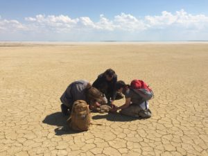
Steffen Zuther of ACBK and Frankfurt Zoological Society with students, taking biological measurements of a calf in the Irghiz region of Kazakhstan in 2016.
Prior to this mass mortality event (MME), the multi-institutional monitoring team had established protocols for health and mortality surveys of the calving aggregations, and regional Kazakh veterinary staff had received some training from the Royal Veterinary College and the Food and Agriculture Organization of the United Nations. This included basic necropsy and outbreak investigation techniques. A manual of standard operating procedures for saiga mortality was undergoing review by the authorities and this ensured that when such a sudden unexpected event occurred, the research team and regional veterinarians were, for the first time, prepared. With a research team in place, the leader, Steffen Zuther, of the Frankfurt Zoological Society and the Association for the Conservation of Biodiversity of Kazakhstan, was able to respond immediately. As soon as he and Sara Wolfs, of the Royal Veterinary College, London, reached the Torgai site, they started recording observations and taking samples. A week later, one of us (Kock) and Mukhit Orynbayev, of the Research Institute for Biological Safety Problems in Gvardeyskiy, Kazakhstan, joined the emergency response team in Tengiz to cover the second event.
As we reported in our paper in Science Advances of 17 January 2018, we “carried out field postmortems and sampling according to an outbreak investigation protocols.” We didn’t attempt to randomly sample the deceased animals for epidemiological analysis— the syndrome was universal and consistent in character—and there weren’t any healthy animals for comparison in the immediate area. Based on later ground and aerial surveys, there were approximately 30,000 individuals unaffected by the outbreak—small herds in isolated areas and a larger herd of older males in the north, away from the calving areas, but the precise location of these herds and their condition was difficult to determine, because the saiga range covers a vast area.
We performed full postmortem examinations on a total of thirty three dead saigas from the Torgai and Tengiz sites of which twenty-four were adult females and nine were calves; four males and five females. We also took samples from twentysix moribund saigas at the Torgai site and four from Tengiz. Samples from dead saigas included blood, feces, gut contents, tissues, and any observed external or internal parasites. Tissue samples from liver, spleen, lungs, kidney, blood, and milk were frozen for later testing for bacterial and viral pathogens—RNA testing for foot-and-mouth disease, bluetongue virus, and peste des petits ruminant virus (which primarily affects goats and sheep), and DNA testing for viruses (sheep pox, Aujeszky, and Visna-Maedi) and blood for protozoa (Theileria, Babesia, Anaplasma, and Coxiella). Whole liver, kidney, brain, rumen, and gut content were taken for plant and toxin studies and tissue mineral analysis from saiga at the Tengiz site to evaluate possible nutrient or micronutrient deficiencies and ecotoxicologies. The distribution of carcasses at the mortality sites showed a remarkably even spacing of approximately thirty to fifty meters between individual adults. This pattern is consistent with animals becoming ill while grazing and dying on the spot. Most died within a few hours of onset of clinical signs. The observed behavior of individual animals changed from running and grazing to general lethargy, hindlimb weakness, and loss of body control, or ataxia, while grazing or standing. Sicker animals were recumbent, some with labored breathing and grunting, frothy fluid or saliva forming around the mouth, and terminal diarrhea, frequently with blood and urine staining of the soil. Postmortems revealed all organs were congested and there were widespread hemorrhages. The on-site veterinarians, Orynbayev and Kock, both agreed that the gross picture was suggestive of haemorrhagic septicaemia (HS).
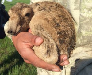
A newborn saiga calf nestles in the arms of a scientist of the joint health monitoring team. Scientists think the saiga’s distinctive nose helps filter out dust kicked up during summer migrations and warms up inhaled air in the winter.
Once we had sent off samples for analysis to Kazakhstan and overseas laboratories, we established how our collaboration should proceed with its formal investigation of the MME. We wanted to ensure that research was carried out in a systematic and structured manner. The research team first listed all the characteristics of the outbreak. We then developed nonmutually exclusive hypotheses—consistent with these characteristics—for the proximate cause of the outbreak. Again, from our Science Advances paper, “These hypotheses were then explored, and refined or discarded, using a combination of review of the English and Russian language literatures (including texts available only in Russia or Kazakhstan), laboratory analyses, expert consultation with field biologists who were present at the time of previous outbreaks, consultation of archived field notes and maps, followup field expeditions, and statistical analysis using a range of data sets of environmental covariates.”
As seasonal migrants—covering a range up to 1,500 kilometers— saiga antelopes of the Betpak-dala rangelands normally have access to greater seasonal food supplies than would be available to resident ungulates. Saigas have a very high investment in reproduction. They produce precocial calves—the largest mass at birth as a proportion of maternal mass of any ungulate—and can reach high numbers in their large ranges (one to two million individuals in Kazakhstan only a few decades ago). Conversely, adverse climatic conditions and a diminished food supply can have a large impact on saigas, affecting reproduction processes, even survival. And in following seasons, the weakened animals may be more susceptible to disease, especially if they are further stressed by extreme weather events. In the past, saigas have contracted parasites and diseases (e.g., foot-and-mouth) transmitted by cattle and sheep, and in December 2016 for the first time Mongolian saigas contracted peste des petits ruminants from domestic sheep and goats with whom they rarely have contact but whose pastures they pass through and whose water sources they share.
After the devastating event in May 2015, we conducted follow-up expeditions that summer and in the spring of 2016 to villages and farms in the region. We didn’t see, nor hear of, any significant, excess livestock deaths or human disease in the vicinity, nor did we find evidence of any large numbers of ill or dead individuals of other wildlife species. From our observations of how the outbreak progressed, it suggested a mode of infection other than horizontal spread between animals. We observed calves suckling until, and after, their mothers died, and then they too died. Delayed death of calves was consistent with disease transmission from the mother’s milk; otherwise, calves would have been infected before or at the same time. Most important, these conditions suggested that there was no aerosol transmission between animals. The only credible pathway was through opportunistic infection of every individual by bacteria latent in the animals already. P. multocida is a commensal bacterium, which can reside in the tonsils of animals, but only causes disease when the immune barrier is breached for some reason, leading to bacterial invasion of the blood and septicaemia. This opportunistic disease is sometimes associated with stress in domestic animals, from sudden adverse weather events such as a tropical storm.
Our preliminary diagnosis, based on the clinical presentation and necropsy, was HS caused by the bacterium Pasteurellosis multocida serotype B. This diagnosis was supported by samples tested at the Research Institute for Biological Safety Problems in Gvardeyskiy, Kazakhstan, at the Royal Veterinary College, and at International Reference Laboratories; Pirbright Institute, UK; and Froedrich Loeffler Institute, Germany.
Based on our hypotheses and on evidence concerning triggers of pasteurellosis from the literature, we tested for any differences between various measures of temperature, precipitation, wind speed, atmospheric moisture, soil moisture, and vegetation greenness in the five– to ten-day period before the die-off and for snow cover in the preceding weeks. As we described in our January paper, “all analyses, average maximum daily relative humidity, average minimum daily temperature, and average maximum daily dew-point temperature showed the strongest positive relationships with the probability of a die-off; in particular, die-off sites were unusually humid, and in 2015, they were unusually warm. Of the Bayesian multivariate models, the strongest associations emerged for the ten-day aggregates of the days preceding the die-off. The final model contained only temperature and humidity metrics. There was a clear threshold association with humidity, with most die-offs occurring at a mean average maximum daily relative humidity of more than eighty percent.” In May 2015, atmospheric moisture was extreme with some of the highest recorded values for that time of year since 1948.
As we concluded in our Science Advances paper, “Recent history suggests that the saiga must be managed with the high probability of future MMEs in mind; this requires investment in preventative measures where possible (for example, livestock vaccination for PPR), strong antipoaching actions so that populations are large and resilient enough to withstand large-scale mortality, and a sustainable landscape-level approach to coexistence between saigas, livestock, and people to enable its migratory lifestyle to continue.
“The fact that P. multocida infection in saigas, as in bovines, appears strongly linked to weather driving high humidity and temperature is of concern going forward, given that a climate change–induced increase in temperature is projected for the region over the short to medium term. “This study illustrates the power of a multidisciplinary, collaborative approach to investigating MMEs, the importance of understanding the contextual factors as well as the proximate cause, and taking a historical, evidence-based approach to investigations. A narrow focus on etiology of MMEs could fail to elucidate causation, where this involves multifactorial interactions between pathogens, hosts, and environment. The scale and nature of this event also point to the need for ongoing scientific and veterinary monitoring of wildlife populations and the need to be prepared for rapid and rigorous responses to disease outbreaks when they occur.”
Richard A. Kock is a wildlife veterinarian, researcher, and conservationist at the Royal Veterinary College, London. Eleanor J. Milner-Gulland is a conservation scientist at the University of Oxford and co-founder of the Saiga Conservation Alliance.
Download the original article here